Dr. Peter Kruger
Patient Resource

Understanding Prolapse Repair

Explore stage 3 prolapse in 3D and see how surgery can restore support at the top, sides and opening of the vagina.

Side view · cutaway
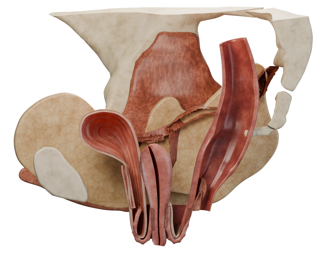
Side cutaway showing the bladder towards the front, uterus in the middle and rectum at the back of the pelvis. On strain.
MovementOn strain
The starting anatomy

Stage 3 prolapse

When you bear down, weakened supports can allow the vaginal walls and the cervix to bulge beyond the vaginal opening. In this example, all three areas descend.

The bladder sits against the front vaginal wall. Both bulge down together.
The uterus moves down and its lower end, the cervix, approaches and passes the opening.
The rectum pushes the weakened back vaginal wall forward into the bulge.

Questions to discuss

How are a stage, a compartment and a level different?

A stage describes how far the lowest point descends when you bear down. In stage 3, it extends more than 1 cm beyond the hymen, the landmark at the opening, and falls short of complete or nearly complete descent. A compartment is the front, top or back region. A level describes a region of support from the top of the vagina down to its opening.

Which repairs would I need?

Your surgeon checks where you feel the bulge, which areas descend and how well each attachment supports the vagina. Your symptoms, preferences and examination guide the plan. Ask which parts of this example apply to you and what other options you have.

What should we discuss about my ovaries?

Removing the uterus and cervix ends the ability to carry a pregnancy. Removing both ovaries before natural menopause also causes surgical menopause. Ask about the benefits and effects of keeping or removing your ovaries and fallopian tubes in your circumstances.

What might change after surgery?

The aim is to reduce the bulge and improve symptoms. Ask about recovery and possible changes in bladder emptying, bowel movements, comfort during sex and urine leakage. Prolapse or leakage can persist or return.

Anatomy credits and sources

The pelvis and surgical changes illustrate one combined operation. The positions and stitches were modelled for teaching. Your examination and discussion with your surgeon guide your care.

Clinical reading

Anatomical artwork

Pelvic structures were adapted from the HuBMAP CCF 3D Reference Object Library v1.2 by Kristen Browne, Heidi Schlehlein, Bruce Herr II, Ellen Quardokus, Andreas Bueckle and Katy Börner. The models were cropped, recoloured, sectioned and deformed for this example. Reference library · Creative Commons Attribution 4.0

Pelvic floor structures were adapted from the Female Pelvic Floor Visualiser by Abbas Talimian, Hana Čechová, Magdalena Jansova and Luděk Hynčík at the University of West Bohemia in Pilsen. The 3D viewer uses Three.js. Pelvic floor dataset · Full credits and MIT licence notices