Overview
Laparoscopic uterosacral ligament suspension is a native-tissue prolapse repair used to restore support to the vaginal apex or cervix. The uterosacral ligaments are strong supportive bands that run from the cervix and upper vagina toward the sacrum. When these supports weaken, the uterus or vaginal vault can descend, causing apical pelvic organ prolapse.
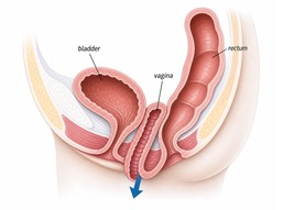
This operation uses sutures, rather than mesh, to reattach the top of the vagina or cervix to the uterosacral ligaments. The laparoscopic approach allows the repair to be performed through small abdominal incisions with direct visualization of the pelvis.
Indications
- Symptomatic apical prolapse involving the vaginal vault, cervix, or uterus
- Vault prolapse after hysterectomy
- Uterine prolapse when a native-tissue suspension is appropriate
- Support of the vaginal apex at the time of hysterectomy or other prolapse repair
- Patients who prefer or are better suited to a non-mesh apical repair
Surgical Technique
- Performed under general anesthesia using small laparoscopic incisions
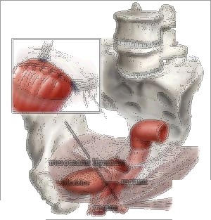
- The pelvis is inspected and the ureters are identified so sutures can be placed safely
- Permanent or delayed-absorbable sutures are placed through the uterosacral ligaments and the vaginal apex or cervix
- The sutures are tied to elevate and support the top of the vagina in a more normal anatomic position
- Cystoscopy is commonly performed during the operation to confirm bladder integrity and ureteral flow
The procedure may be combined with hysterectomy, anterior repair, posterior repair, or an anti-incontinence procedure when clinically indicated.
Benefits
- Uses native tissue without permanent mesh
- Restores support to the apical compartment, which is central to durable prolapse repair
- Maintains a more natural vaginal axis compared with some vaginal suspension approaches
- Laparoscopic visualization can help with ureter identification and precise suture placement
- Recovery is generally faster than with open abdominal surgery
Recovery
- Hospital stay: typically same day to 1 night, depending on combined procedures
- Temporary urinary catheter may be used after surgery
- Light activity can usually resume within 1-2 weeks
- Avoid heavy lifting and strenuous exercise for approximately 6 weeks
- Full recovery is typically 4-6 weeks, longer if combined with more extensive surgery
- Sexual intercourse is usually avoided until postoperative review confirms adequate healing
Risks and Success Rates
Reported success rates for uterosacral ligament suspension are approximately 80-90%. As with any prolapse surgery, recurrence can occur over time, and another vaginal compartment may develop prolapse in the future.
Potential risks include:
- Ureteric kinking or injury — the ureters run close to the uterosacral ligaments; cystoscopy is commonly used to confirm ureteral flow
- Urinary tract infection — more common when a catheter is used
- Voiding difficulty — usually temporary, but may require short-term catheter use
- Bleeding or hematoma — uncommon, but possible with any pelvic surgery
- Infection — wound, bladder, or pelvic infection
- Pain with intercourse — uncommon, but possible with scarring or vaginal narrowing
- Buttock or pelvic pain — usually short-term when it occurs
- Injury to bladder, bowel, blood vessels, or nerves — uncommon but possible
- Venous thromboembolism — rare, but a recognized risk after pelvic surgery